


SWOLLEN RIGHT FOREARM IN A YOUNG MALE STUDENT
INTERESTING CASE
Submitted by H Thomas, CJ Evans, RM Evans, Withybush Hospital, Hywel Dda Heath Board.
Clinical history :
An 18 year male student was referred by his GP for an Ultrasound examination. He presented with a three month history of a swelling of his right elbow/forearm. The clinical history given on the referral was :
“Intermittent swelling right forearm, ? RSI worse on typing /keyboard (student)” and had been booked as a routine appointment.
Ultrasound examination of the elbow and proximal forearm was performed, a series of images are presented below :
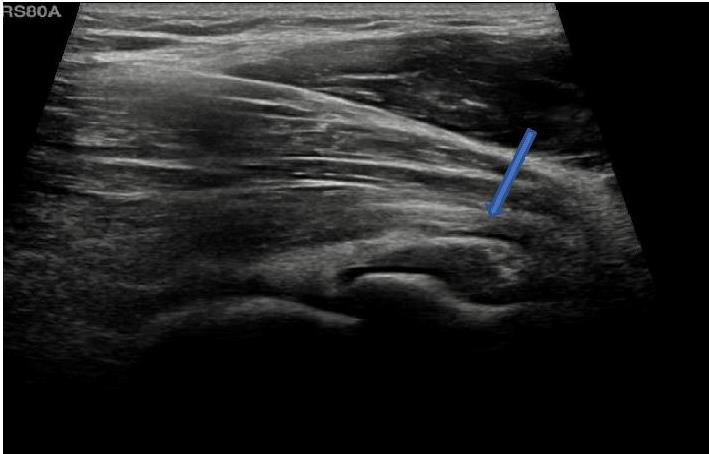 |
Image A : Longtitudinal image : small effusion seen anterior to the radial head (arrow). |
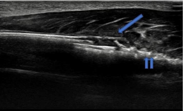 |
Image B : Longitudinal image of dorsal aspect of proximal right ulna : demonstrates a soft tissue mass (arrowed) elevating the periosteum at the margin of an area of permeative cortical destruction (double arrow). |
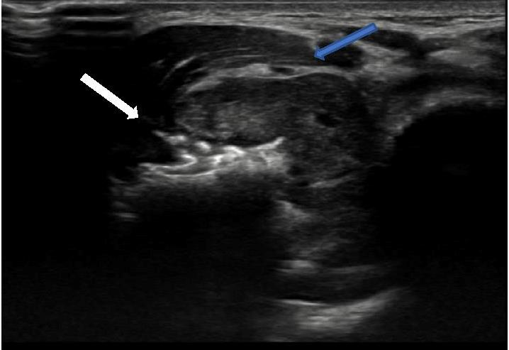 |
Image C : Transverse image proximal ulna : spiculated tumour (white arrow) arising from right ulna with associated soft tissue mass, note preservation of fat plane between tumour & overlying muscle (blue arrow). |
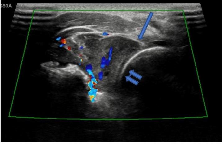 |
Image D : Transverse image of dorsal proximal forearm, demonstrates vascularity of tumour. Arrow demonstrates tumour abutting radial head of proximal right radius (double arrow). |
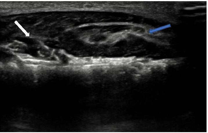 |
Image E: Longitudinal image showing cortical destruction with spiculated outline due to aggressive bone lesion (white arrow) , lobular soft tissue tumour elevating overlying muscle compartment (blue arrow). |
 |
Image F : Transverse image proximal right ulna :highlighting sunburst like periosteal reaction/cortical profile (arrow), typical of a primary bone tumour. |
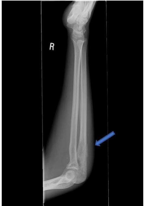 |
Image G : Lateral radiograph right radius and ulna – periosteal elevation (Codman triangle) adjacent to an area of permeative bone destruction in proximal ulna . |
What is the probable diagnosis based on the imaging findings?
| To see the diagnosis, please click here |