


An unusual finding for left iliac fossa pain
CASE OF THE MONTH
Submitted by : Georgiana Zamfir1, Peter Cantin2, 1Peninsula Radiology Academy/ 2Derriford Hospital, Plymouth
Presentation
A 61-year-old female patient was referred for an abdominal ultrasound to investigate 3 day history of left iliac fossa pain. On further assessment, the pain was described as being intermittent and exacerbated by movement. There were no urinary symptoms and no change in bowel habits. The patient was otherwise fit and well and had a previous cholecystectomy.
Representative sonographic images of the abdominal scan are shown below:
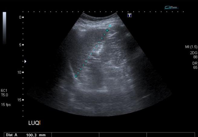 |
Figure 1. Longitudinal image of the spleen |
 |
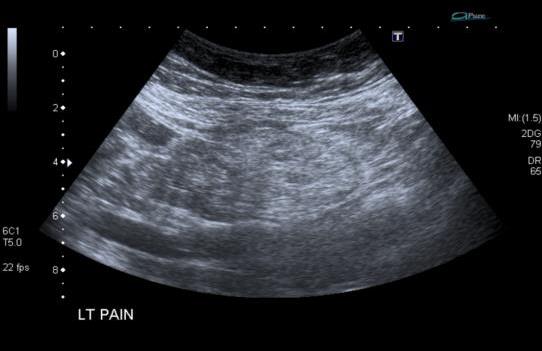
Figure 2. Longitudinal image of the left kidney |
 |
Figure 3. Transverse image through the left upper quadrant |
 |
 |
|
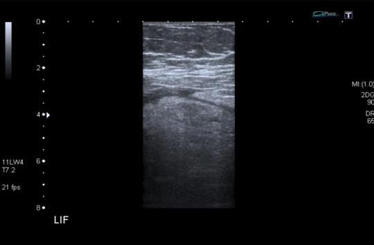
Figure 4. Targeted longitudinal sections over the LIF |
What is the most likely diagnosis and differential diagnosis?
To see the diagnosis and differential diagnosis, please click here