


Ultrasound guided core biopsy of a glottic cancer
INTERESTING CASE
Submitted by : JJ Thomas and RM Evans. Withybush Hospital, Hywel Dda University Health Board
Clinical History :
A 74 year old lady presented with progressive sore throat and hoarse voice.
Image Findings :
Contrast CT demonstrated a soft tissue mass involving the left vocal cord, extending into the left and right paraglottic fat (Figure 1).
 |
Figure 1. Axial CT showing soft tissue mass as described above. |
Direct laryngoscopy was attempted but biopsy of the lesion was not possible. The overlying mucosa appeared to be benign (Figure 2).
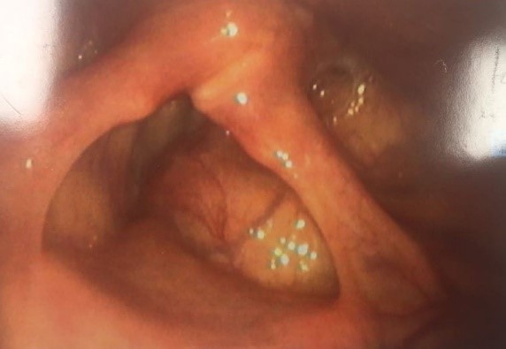 |
Figure 2. Benign mucosal appearance on endoscopy. |
A provisional diagnosis of a possible laryngocele was made, given the normal mucosal appearances. Despite repeated attempts, it was not possible to direct a laryngoscope into the laryngeal ventricle to assess the larynx appropriately.
Her symptoms persisted and progressed over the following 6 months. Another CT was performed, showing progression of the lesion, with further invasion into the right paraglottic fat (Figure 3).
 |
Figure 3. Axial CT- contralateral extension of mass into the paraglottic fat (white arrow). |
There was no invasion into thyroid cartilage nor into the hypopharynx. Again, biopsy via laryngoscopy was not possible.
A decision was made to proceed to Ultrasound to see whether (a), any lesion could be seen and (b), whether a biopsy was possible.
The lesion was readily visualized on ultrasound (Figures 4 and 5).
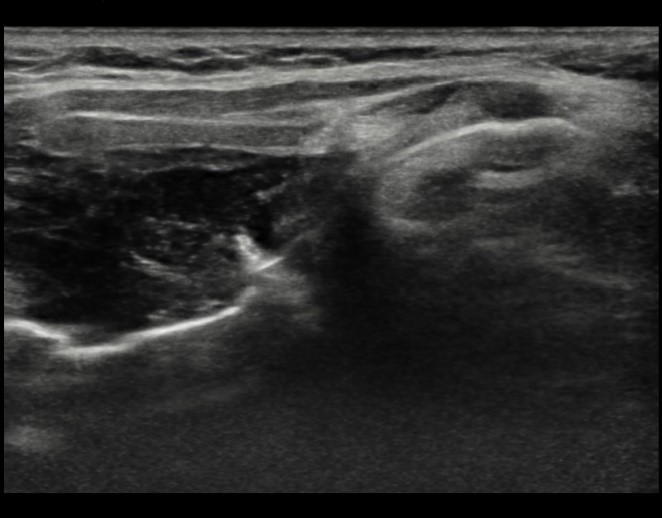 |
Figure 4. Longitudinal image through larynx.
|
|
Figure 5. Transverse image |
What is the probable diagnosis based on the imaging findings?
| To see the diagnosis, please click here |